
Журнал «Боль. Суставы. Позвоночник» 2 (18) 2015
Вернуться к номеру
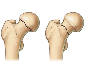
The Indicators Bone Metabolism in Fractures of Trochanteric Areas of Femur in Patients with Type 2 Diabetes
Авторы: Shymon V.M., Stoyka V.V., Sheregiy A.A., Shymon M.V., Slyvka R.M. - Uzhgorod National University, Uzhgorod, Ukraine
Рубрики: Ревматология, Травматология и ортопедия
Разделы: Медицинские форумы
Версия для печати
Статья опубликована на с. 95-96
Introduction. Fractures of the trochanteric area of the femur is an actual problem. There are the peculiarities regeneration of fractures of the trochanteric area of the femur in patients with type 2 diabetes. This is due to the peculiarities of osteoreparation background of type 2 diabetes. In recent decades was conducted a large number of studies to determine changes in bone in patients with type 2 diabetes. The available data are very different. Thus, according to different authors an increase in bone mineral density (BMD), normal density and decrease in BMD compared with controls can be observed. In a meta–analysis of the observed experiments–LMa and co–authors (2012) noted that these differences may be related to differences in the design of experiments methodology for determining BMD, samples of patients and the presence of complications. We believe that individual indicator BMD may not fully reflect the osteoreparative processes. Comparison of data ultrasound mineral densitometry and hormonal balance will help to understand the changes in bone metabolism in patients with type 2 diabetes and help to find ways to prevent complications in the treatment of fractures.
Aim. To study changes in bone metabolism in fractures of trochanteric area of the femur in patients with type 2 diabetes.
Materials and methods. In the period since 2012 to 2015 were examined 42 patients who were hospitalized in the clinic at the department of general surgery UzhNU about fractures of the trochanteric area of the femur. There were 34 injured women, men — 8. Age composition ranged from 48 to 79 years, average age — 67 years. Before trauma patients had active lifestyle.
The main group consisted of 19 patients with type 2 diabetes. Body mass index (BMI) was 29.4 (25.7–34.2) kg/m2, level of glycosylated hemoglobin (HbA1c) — 9.6 (7.7–11.3) %. Part of patients (n = 13) took sulfonylurea drugs 10.5 (7–10.5) mg/day, metformin — 1.5 (1–1.5) g/day, and other (n = 5) received combined treatment with insulin. The average duration of the disease diabetes was 8 years old. Newly diagnosed diabetes was 1 case, in 4 patients disease duration was less than 5 years, 7 patients — from 5 to 10 years, 7 patients — more than 10 years. Among the complications of diabetes was more frequent diabetic micro and macro angiopathy limbs — 9 people, diabetic retinopathy — 5 people, diabetic neuropathy — 4 people. Cardiovascular diseases were observed in 13 people.
The control group consisted of 23 patients with fractures of the trochanteric area of the femur in which rates of sugar of blood serum and glycated hemoglobin does not exceed the norm. BMI was 28.9 (25.3–33.9) kg/m2. Both groups were comparable in age, sex, severity of the general condition and the nature and methods of fracture surgery.
On all patients were performed laboratory and instrumental clinical study determined the level of calcium, phosphorus, hydroxyvitamin 25(OH)D, alkaline phosphatase, parathyroid hormone (PTH) and osteocalcin.
To determine BMD ultrasonic densitometry was performed in three standard parts of the skeleton (lumbar, proximal femur, forearm). Were estimated bone mineral density L1–L4 spine, proximal femur and distal forearm. BMD assessment was performed according to WHO re–commendations Z– and T–criteria. In women during menopause and men over 50 years using T–test data in accordance with the interpretation of densitometric WHO classification (rate from 2.5 to 1, osteopenia from –1 to –2.5, osteoporosis from –2.5 SD and below).
Results and discussion. In people with diabetes type 2 breach phosphorus–calcium balance may be at different stages of the disease, and many researchers indicate normal or slightly reduced levels of calcium and phosphorus in the blood of patients, which coincides with our data. In the experimental group levels of calcium was 2.13 ± ± 0.03 mmol/l, and phosphorus — 1.05 ± 0.04 mmol/l. In the control group levels of calcium was 2.37 ± 0.03 mmol/l, and phosphorus — 1.28 ± 0.04 mmol/l. The same applies to hydroxyvitamin 25(OH)D, the level of which was in the normal range in both groups. Alkaline phosphates’ level in experimental group was 128.70 ± 0.04 IU/l, in the control group — 156.30 ± 0.04 IU/l.
Important role in the regulation of bone formation and osteoreparation pay parathyroid hormone. In the experimental group was marked higher levels of parathyroid hormone than in the control group, and was 58.08 ± 2.70 pg/ml and 41.55 ± 1.90 pg/ml, respe–ctively.
Nowadays it is admitted that the most informative indicator of bone growth is the level of osteocalcin, which is synthesized by osteoblasts. In patients with type 2 diabetes osteocalcin level was 19.92 ± 0.80 ng/ml, in control group — 32.87 ± 0.90 ng/ml.
In the study of data of densitometry in three locations (lumbar, proximal femur, forearm) reduction in bone mass (T < –1) detected in most patients, with those in the expe–rimental group it was more pronounced than in the control group. In the experimental group prevailed patients with a diagnosis of osteoporosis (T < –2.5) in the control group prevailed patients with osteopenia.
When comparing the densitometric measurements in three standard points were observed that in the experimental group osteopenic syndrome most often seen in the proximal femur (78.9 %), whereas in the control group were most pronounced changes in the lumbar spine (73.9 %).
Conclusion. The severity and duration of type 2 diabetes affect the bone metabolism and cause leakage osteoreparation in patients with fractures of the trochanteric area of the femur through changes in mineral and hormone balance.
The possibility of less reversible changes in bone tissue on a background of type 2 diabetes poses the need to address the issue of early diagnosis and treatment of abuse of bone metabolism in this group of patients.