
Журнал «Боль. Суставы. Позвоночник» Том 9, №4, 2019
Вернуться к номеру
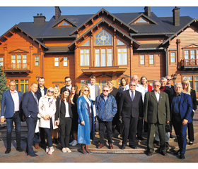
Healthcare of Bone and Joint Diseases (International Scientific-Educational Conference, October 21–22, 2019, in Kyiv, Ukraine)
Авторы: N. Kuprinenko
Рубрики: Ревматология, Травматология и ортопедия
Разделы: Медицинские форумы
Версия для печати
During the recent five years, holding the international meetings for scientists and practitioners, interested in the problems of osteoporosis and other bone and joint di–seases, has become a good tradition. On October 21–22, 2019, the International Scientific–Educational Confe–rence «Bone and Joint Diseases and Age» was held in Kyiv (Ukraine). This event was organized by the Ukrainian Association of Osteoporosis, Ukrainian –EVIDAS Division, D.F. Chebotarev Institute of Gerontology by the NAMS of Ukraine, in collaboration with the National Academy of Medical Sciences of Ukraine, National Society of Ge–rontology and Geriatrics of Ukraine, Ministry of Health of Ukraine, Ukrainian Association of Orthopedic Traumatologists and Association of Rheumatologists of Ukraine. The Organi–zing Committee of this annual International Conference, as always, was headed by Professor Vladyslav Povoroznyuk, President of the Ukrainian Association of Osteoporosis. The conference had a wide outreach: many foreign lecturers, a wide range of to–pics, and multitudes of participants. 18 presenters from various countries of Europe, the USA and Kazakhstan, as well as nume–rous Ukrainian lecturers, made 44 reports, reviews and lectures. The simultaneous interpretation of the lectures, presentations and discussions was provided, so that all the participants of the symposium could get comfor–tably immersed into the new and useful expertly information.
The scientific program of the symposium opened with the lecture by the President of the Ukrainian Association of Osteoporosis, Prof. Vladyslav Povoroznyuk (Chebotarev D.F. Institute of Gerontology by the NAMS of Ukraine, Kyiv, Ukraine). He was reporting on osteoporosis and musculoskeletal diseases in Ukraine and presenting activities and achievements of the Ukrainian Association of Osteoporosis and Ukrainian EVIDAS Division. Along with that, Prof. V. Povoroznyuk also presented the Ukranian version of FRAX, algorithm measuring a 10–year probability of hip and major osteoporotic fractures in patients aged 40 years and older. As of June 2016, the FRAX questionnaire could be used in the Ukrainian language, an outstanding feat by the staff of the Ukrainian Scientific–Medical Center of Osteoporosis and D.F. Chebotarev Institute of Gerontology by the NAMS of Ukraine. The Ukrainian model of FRAX relies on a series of epidemiological studies, conducted in the recent years by the members of the Ukrainian Association of Osteoporosis and Ukrainian–Scientific Medical Center of Osteoporosis and endorsed by the Ukrainian Association of Traumatologists and Orthopedists. In October 2016, the Ukrainian model had been launched on the official FRAX website. The Ukrainian model of FRAX was the first national model of 10–year fracture probability measurement, with risks of major osteoporotic fractures and hip fractures in mind. It is based on the original FRAX methodology, developed by the World Health Organization Collabora–ting Centre for Metabolic Bone Diseases. Afterwards, Prof. V. Povoroznyuk was holding master classes on back pain and cerebral and cardiac manifestations of cervical and thoracic osteochondrosis.
Prof. Neil Binkley (University of Wisconsin School of Medicine and Public Health in Madison, Wisconsin, USA) presented three reports. His first report was on sarcopenia and fracture prevention in the elderly. The urgency of this problem is underlined by the recent statistical prognosis, accor–ding to which by 2050 the elderly population in Europe would amount to 34 %, in the North America — to 28 %, in the Latin America — to 26 %, in Asia — to 25 %, in Oceania — to 23 %, and in Africa — to 9 %. It is a well–known fact that fracture risk increases with age. Many osteoporosis–related fractures, including over 90 % of hip fractures, occur due to falls. Falls become common with advancing age: about every one of three adults aged 65 and > 40 % over age 75 fall each year. It is worthy of noting that only 44 % of women (and 21 % of men) who sustained non–vertebral fractures had “osteoporosis” diagnosed by the BMD. Various trials confirm the significant association between sarcopenia and osteoporosis in a large sample of hip–fracture women and support preventive strategies and treatment options for sarcopenia and osteoporosis targeting both bone and muscle (Di Monaco et al., 2011). However, treating bone and muscle is not enough. Obesity as a key factor of increasing fracture risk also should be taken into account. The global longitudinal study of 60,393 women aged ≥ 55, who had been followed for 2 years, demonstrated that obesity was not protective against fractures in postmenopausal women and was associated with an increased risk of ankle and upper leg fractures (Compston et al., 2011). This is where the concept of sarcopenic obesity comes from. Rather than focusing on a single component, i.e., osteoporosis, sarcopenia or obesity, it is important to combine clinical factors, thereby enabling an improved identification of older adults at risk. Such a combination may be referred to as ‘dysmobility syndrome’. The above–mentioned umbrella term allows consideration of other things that might cause difficulty in walking or increase falls risk, e.g., balance, osteoarthritis, neuropathy, etc. (Binkley et al., 2013). As far as the treatment is concerned, the role of exercise/physical therapy in falls risk reduction has been proved, as well as the role of protein, taken in the amount of 1.2–1.5 g/kg daily, calcium and Vitamin D.
In his second report Prof. Neil Binkley elaborated on the vitamin D deficiency and insufficiency. He observed that in the USA the prevalence of vitamin D deficiency was comparable to obesity, hypertension and hyperli–pidemia. The question “How to define “low” vitamin D status” was still moot. At the moment, to assess the status of vitamin D, the level of 25(OH)D was measured, but it has strikingly small values and varies according to more than 1 factor (calcium intake, PTH, etc.); its measurement considered variable (DEQAS Data, 2018). Thus, it seems likely that we need “vitamin D panels” or ratios, including 25(OH)D, PTH, 3–epi 25(OH)D, cholecalci–ferol, 24,25(OH)2D.
Together with Prof. Didier Hans (Center of Bone Di–seases, Lausanne University Hospital Switzerland, in Lausanne, Switzerland), Prof. Neil Binkley presented a 6–step outline of Trabecular Bone Score (TBS)’s practical clinical use. TBS is acknowledged worldwide as an independent parameter for fracture prediction, providing fracture risk information independent of BMD and CRF. The main steps of using TBS include checking image quality (edge detection and artifacts), individual patient’s characteristics of bone microarchitecture, determination of bone health categories (normal bone, osteopenia, and osteoporosis) and the risk of fracture, choosing the treatment options and, afterwards, lowering the fracture risk zone. It is worthy of no–ting that if the patient were out of the bone–based risk zone, it did not mean that he/she had no risk of fracture anymore, rather that there was a need to reassess treatment, taking into account the residual risk.
The report by Diane Krueger (University of Wisconsin–Madison, USA) examined the DXA testing quality issues worldwide, as well as the appropriate DXA acquisition and analysis techniques and artifacts and unaccep–table DXA scans. Diane Krueger enumerated the rules for the correct spine, femur and forearm positioning on DXA scans for a proper analysis. The spine must be positioned straight and in middle of scan field, it must be ascertained that bone mar–kers were on the edge of bone and intervertebral markers were in the disc space, vertebral numbering was correct (for example, the L5 marker looked like a bow–tie); the femur shaft must be straight and, ideally, slightly internally rotated, it must be ascertained that bone edges were correct, femoral neck region of interest correctly placed, pelvis or greater trochanter were not included in the scan; forearm must be straight and centered, distal end of radius and ulna must be placed in the scan field, and it must be ascertained that the bone edges were correct and one–third radius region of interest correctly placed.
Prof. Andrea Ildikó Gasparik (Association for Prevention of Osteoporosis in Romania, Bucharest, Romania) told about the health technology assessment (HTA), increasingly used in the field of osteoporosis and potentially able to help decision makers efficiently allocate health–care resources. She highlighted major developments in the economic evaluation: incorporation of me–dication adherence into the pharmacoeconomic ana–lyses, use of FRAX in the health economics, use of micro–simulation models, which were beginning to supplant cohort models in the HTA, direct comparison of the active comparators in the absence of randomized controlled trials, use of indirect treatment comparisons and network meta–analysis to provide useful evidence for selecting the best option.
Prof. Heinrich Resch (Medical School by Sigmund Freud University, Center of Rheumatic and Bone Di–seases Rheumazentrum Oberlaa, Vienna, Austria) reported on the glucocorticoid–induced osteoporosis. It is a well–known fact that glucocorticoids have a direct effect on bone metabolism, contributing to the osteoporosis and fractures. The fractures occurred in 30–50 % of patients taking a chronic glucocorticoid therapy. Glucocorticoid u–sers (with prednisone equivalent doses of ≥ 2.5 mg/day for > 3 months) had the following risks compared to the non–users: up to 2.3–fold higher hip fracture risk, up to 5.2–fold higher vertebral fracture risk, both independent of underlying disease, age, and gender. Additionally, the combined effect of high doses of glucocorticoids used continuously over long terms, might increase the relative risk of hip fractures by 7 times, and vertebral fractures by 17 times. Glucocorticoid effect had two phases: a rapid phase, or the phase of osteoblast suppression and synchronous increased resorption (12 % BMD loss within the first 3–12 months); and a slow phase, or the phase of slow resorption, however attended by the suppressed bone formation (2–5 % BMD loss/year). The registered therapy of glucocorticoid–induced osteoporosis involved: for postmenopausal women — alendronat (10 mg/day), risedronat (5 mg/day), zoledronat and teriparatid; in males — alendronat (10 mg/day), teriparatid and zoledronat. In RCTs of glucocorticoid–induced osteoporosis, teriparatid had shown a significant potential for reduction of vertebral fractures in comparison to alendronat. Denosumab (60 mg) taken for over 1 year was associated in adults with an increased fracture risk and glucocorticoid–induced osteoporosis.
The topic of secondary osteoporosis was continued by the report of Prof. Mario Rui Mascarenhas (Lisboa, Portugal). That diagnosis was suspected in the event of fracture for premenopausal woman and for men younger than 70–75 years, in the event of fractures for the young persons, in the event of the frequently occurring fractures, repeated fractures at the unusual skeletal sites, in the event of dissociation between the fracture site and age of the patient (namely, hip fracture occurring before 70 years), in the event of DXA –Z–score < –1.5 or < –2.0 SD, in the event of BMD reduction despite antiosteoporotic therapy. The diagnostic tests for secondary osteoporosis included the proteinogram, blood calcium, phosphorus and urinary calcium alkaline phosphatase and iron le–vels, the assessment of kidney function, liver function and thyroid function: TSH (FT3 and FT4), gonadal function: testosterone, estradiol, LH/FSH (PRL), iPTHi, Vitamin D, 24h urinary cortisol, CRP (US), anti–gliadin AB and/or duodenal biopsy.
Prof. Juraj Payer (Comenius University Faculty of Medicine in Bratislava, University Hospital of Bratislava, Slovak society for osteoporosis and metabolic bone di–seases, Bratislava, Slovakia) summarized the key issues in the field of premenopausal osteoporosis. Most prevalent causes of secondary osteoporosis in pre–menopausal wo–men were: glucocorticoids, estrogen deficiency (premature ovarian failure, anorexia nervosa), pregnancy and lactation associated osteoporosis (gastrointestinal diseases, celiac disease), intestinal bowel di–sease, malabsortion, certain medications, endocrine di–seases (primary hyperparathyroidism, hyperthyroidism, GH disorders), alcohol abuse, osteogenesis imperfecta. Low BMD in the premenopausal women was not associated with the same risk of fracture as in postmenopausal women. Results from densitometry alone might be used for diagnosis of premenopausal osteoporosis; however, additional tools such as TBS could help diagnose (exclude) secondary osteoporosis. Most cases of premenopausal osteoporosis were caused by the underlying se–condary disease and associated with a poor bone quality. General recommendations about the diet and physical activity should be delivered to all the premenopausal women. A specific antiporotic treatment should be considered on a case–by–case basis.
Prof. László Hodinka (National Institute of Rheumatology and Physiotherapy, Budapest, Hungary) paid attention in his report to the role of cytokines in pain proces–sing. He observed that the pain decrease in the rheumatoid arthritis JAK–inhibitor study had been more pronounced than expected from extrapolation of the proportional anti–inflammatory effect on CRP, erythrocyte sedimentation and swollen joint count. The difference had been attributed to the direct analgesic effect of the cytokine–signaling JAK–STAT pathway. Furthermore, an enhanced brain blood flow observed in the functional MRI study of rheumatoid arthritis patients was associa–ted with an activated state of the pain processing cerebral are–as, but gradually decreased in patients well responding to the TNF inhibiting anti–cytokine therapy. Cytokine signaling targeted small mole–cular therapy (Janus kinase inhibitors) and anti–cytokine biological, as IL–6 receptor inhibitor tocilizumab had decreased rheumatoid arthritis pain and had improved functional activity.
The topic of musculoskeletal manifestation of hyperparathyroidism was discussed in the report by Prof. Janusz E. Badurski (Polish Foundation of Osteoporosis, Bialystok, Poland). The classical hyperparathyroidism was associated with: osteopenia, osteoporosis, osteitis fibrosa cystica, bone cyst, brown tumors of long bones, hypercalciuria, nephrolithiasis, nephrocalcinosis, myopathy, a sense of weakness, peptic ulcers, pancreatitis, vascular calcification, arthropathy. As far as the influence of PTH on cartilage is concerned, the PTH did not interfere with a proliferation but with an inhibited chondrogenic differentiation of chondroprogenitor cells. The PTH reduced human cartilage in vitro regeneration, reduced DNA and glycosaminoglycan content, negatively influenced cartilage quality, while theintermittent recombinant PTH inhibited cartilage formation. Chondrocalcinosis was associated with ageing, osteoarthritis, hypomagnesaemia, hemochromatosis, hypophosphatasia, hypercalcaemia and hyperparathyroidism. The pseudogout syndrome (intermittent attacks of arthritis associated with chondrocalcinosis) had been suggested to be the diagnostic clue of hyperparathyroidism or a complication after parathyroidectomy that induced hypercalcaemia on or after the second day after surgery (in one or more joints, often involving the knee). Neuromuscular involvement in patients with hyperparathyroidism manifested itself in a generalized weakness and easy fatigability, 52 % repor–ted paresthesias and muscle cramps, 29 % — loss of pain sensation or loss of vibratory sensation with diminished reflexes. The muscular weakness in patients with hyperparathyroidism had not been caused by neuromuscular transmission. No typical myopathy had been observed. In 44 %, the bone pain and proximal myopathy had been registered; 50 % had had bone fracture. Impaired muscle function might contribute to an increased fracture risk independent of bone mineral density. At the end of his report, Professor Janusz E. Badurski emphasized that some musculoske–letal symptoms observed in PHPT patients could be quite unusual and might represent diagnostic dilemmas to the practicing rheumatologist, endocrinologist, orthopedist and neurologist.
Prof. Nenad Prodanovic (University Clinical Center, Banja Luka, Bosnia and Herzegovina) spoke about key aspects of rheumatoid arthritis (RA) and bone metabolism association. He observed that the greatest loss of bone mass occurred at the early stages of RA and were significant du–ring the first two years of the disease onset. Measurement of bone density should be perfored correctly after the diagnosis and followed up after one year in all patients with rheumatoid arthritis. Continuous administration of corticosteroids, in addition to the disease activity, led to osteoporosis and the use of antiosteoporotic drugs was necessary. The Z–score was considered an isolated risk factor for osteoporosis and an important parameter influencing decisions on the modality of osteoporosis treatment in RA. Osteoporosis associated with RA represented a higher relative fracture risk than osteoporosis of another etiology. The osteoporosis management strategies in people with RA included a daily calcium intake of 1000 mg for men and women up to the age of 50. Women over 50 and men over 70 should increase their intake to 1200 mg daily. As to Vitamin D, the recommended intake was of 600 to 800 IU each day. Medication treatment included bisphosphonates, estrogens, selective estrogen receptor modulators, PTH analog, PTH–related protein analog, RANKL inhibitor, tissue–selective estrogen complex.
Chokan Baimukhamedov (Medical Center of Joint Diseases, Shymkent, Kazakhstan) presented the report about an early rheumatoid arthritis in the elderly. Elderly onset RA (EORA) targets patients who developed RA after the age of 60. EORA is characterized by a lower frequency of rheumatoid factor (RF) positivity, a more frequent involvement of large joints, a higher frequency of acute onset with constitutional symptoms (fever, weight loss and fatigue), a higher frequency of comorbidities and geriartic syndromes, overlapping with other syndromes such as polymyalgia rheumatica (PMR) and remitting seronegative symmetrical synovitis with pitting oedema (RS3PE). Chokan Baimukhamedov presented the Criteria EORA Diagnostic Score, which included clinical and laboratory symptoms, X–ray criteria and negative criteria.
The topic of bone health in childhood cancer was discussed by Prof. Gerold Holzer (Medical University of Vienna, Austria). It had been observed that during the recent 30 years changes in the treatment of children and adolescents with cancer had led to substantial improvements in survival, with a 5–year survival rate of childhood cancer growing up to 80 %. Ho–wever, childhood cancer survivors (CCSs) had the multifactorial bone fragility. Particular attention during cancer treatment should be paid to reduce the impact on future adult bone health. The most commonly used chemotherapeutic agents in childhood cancer were associated with a reduced BMD. In the children treated for acute lymphoblastic leukemia, low doses of methotrexate suppressed osteoblast activity and stimulated osteoclast recruitment. Higher cumulative doses of methotrexate had been associated with a greater incidence of osteopenia. Furthermore, methotre–xate and cisplatin were nephrotoxic and might cause skeletal abnormalities. Ifosfamide could damage renal tubular function and induce the Fanconi syndrome. In the long–term survivors of acute lymphoblastic leukemia, ifosfamide had been found to negatively affect BMD. IOF Working Group recommended BMD evaluation (grade evidence: mode–rate), followed by laboratory exams, in order to assess bone metabolism, renal function and factors indu–cing secon–dary osteoporosis. When low BMD was reported –(juvenile osteoporosis), Z–score < –2 or T–score < –2.5 and/or fragi–lity fractures and/or chronic use of glucocorticoids, antiresorptive treatments (bisphosphonate) should be taken into consideration. Also, if necessary, correction of endocrine alterations or other modifiable risk factors of impaired bone quantity/quality should be evaluated.
Pawel Pludovski (The Children’s Memorial Health Institute, Warsaw, Poland) presented the preliminary results of Polish National Grant study of idiopatic infantile hypercalcemia (IIH). It had been revealed that –CYP24A1 mutations caused majority of true IIH; 25(OH)D and 1,25(OH)D concentrations were often increased; bone mass, bone density, bone strength, bone geometry were normal; muscle mass, fat mass and their interrelationships were normal. Compliance to the restricted diet, the vitamin D supplement use and sun avoidance seemed all crucial to limit clinical complications. However, in the pediatric setting the healthcare providers should be on the lookout for the hypercalcemic individuals who might be hypersensitive to vitamin D.
Jerzy Konstantynowicz (Medical University of –Bialystok, Poland) in his report touched on vitamin D in adolescence. The associations between vitamin D deficiency and morbidity in adolescents had been evaluated in various studies. It has been proven that hypovitaminosis D was common in obese ado–lescents and conferred the risk of diabetes/insulin resistance. Decreased 25(OH)D in adolescents was associated with hyperglycaemia and metabolic syndrome, CVD risk and hypertension, independently of obesity. That is why the healthcare provi–ders aim is to put vitamin D back in children and adolescents. Current recommendations promoting the intake of 10–15 µg/day might be helpful in preventing the vitamin D deficiency; however, higher doses (20–50 µg/day) might be necessary to ensure the optimal level. In cases of severe vitamin D deficiency (< 10 ng/ml), the do–sage of 50 000 IU once weekly through 8 weeks should be prescribed. Children and adolescents with obesity should be administrated 1200–2000 IU/day (30–50 µg/day) throughout all seasons.
The Ukrainian — Austrian summit for bone and joints diseases also took place in the framework of the Confe–rence. The scientists from these countries presented data of their studies on sarcopenia, hypophosphatasia, role of micro–RNAs in the diagnosis of osteoporosis, secondary osteoporosis attending the internal diseases, evaluation of BMD and TBS in the male population, risk of falls in the gerontological practice.
The final chord of the International Scientific–Edu–cational Conference “Bone and Joint Diseases and Age” was the expanded meeting dedicated to the 65th anniversary of Professor Vladyslav Povoroznyuk, Pre–sident of the Ukrainian Association of Osteoporosis, President of the Ukrainian Association of Menopause, Andropause and Bone and Joint Diseases, President of the Ukrainian EVIDAS division, Head of the Clinical Physiology and Pathology of Locomotor Apparatus Department of the D.F. Chebotarev Institute of Geronto–logy by the NAMS of Ukraine. The editorial team of our journal echoes the warm words and wishes that were addressed to Professor Vladyslav Povoroznyuk. We wish Vladyslav Povoroznyuk new creative ideas, success, health and all the best!
Prepared by Nataliia Kuprinenko
Dear Vladyslav, we are in Kyiv airport on our way home. Thank you for an excellent meeting and the outstanding hospitality!
Neil Binkley and Diane Krueger
Dear Vladyslav, I hope that the entire meeting and your anniversary came true like you wished. On behalf of me and Andrea, I would like to express many thanks for your kindness and friendship, and a really exciting stay in such a beautiful city.
Once more, all the best
Yours, Juraj Payer
Dear Prof. Povoroznyuk, thank you again for the invitation to the Kiev Meeting. As I said several times I always like to travel to the Ukraine and meet you and all the other colleagues I got to know throughout the many years. Happy birthday again — ad multos annos. Looking forward to meeting you soon again.
All the best,
Gerold Holzer
Dear Vladyslav, you have a lot of reasons to be happy, after a perfectly organized conference!
It was a pleasure to hear words of praise and gratitude from your associates, academics.
I personally thank you, on behalf of my young associates, and on my own behalf, for the invitation to participate in the Conference.
Best regards from Banjaluka
Yours, Nenad Prodanovic
Dear Vladyslav, I am writing in order to let you know how I did enjoy being those days in your company and at the big and extraordinary Congress you organized. Fantastic! I also thank you for your fantastic hospitality and letting us know more about the history and culture of your beautiful country, Ukraine, that you make us love more and more. And also the Ukrainian people that are so kind to us. I want to let you know that whenever you need me, please just let me know.
With my very best greetings
to your family, Ukrainian friends and you,
Mário Mascarenhas
Dear Professor Povoroznyuk, dear Vladyslav, thank you very much for your kind words. It is always a great pleasure, for ESCEO and for myself, to support your prestigious event. I am very happy if my letter has been perceived as appropriate. I really hope to have the privilege of visiting you, in a near future, in your beautiful country.
Warmest regards,
Jean-Yves Reginster
Dear Vladyslav, thank you again for the opportunity to participate at and to contribute to the excellent conference. The presentations were of a great scientific value and the company and accommodation were extremely friendly, the logistics was also first class. I wish similar successes for the future and good health after the 65 yr jubilee for the forthcoming years.
With a continuing warm friendship,
László Hodinka
/269-1.jpg)
/270-1.jpg)
/271-1.jpg)
/272-1.jpg)