Інформація призначена тільки для фахівців сфери охорони здоров'я, осіб,
які мають вищу або середню спеціальну медичну освіту.

Підтвердіть, що Ви є фахівцем у сфері охорони здоров'я.

Журнал «Боль. Суставы. Позвоночник» Том 10, №2, 2020

Вернуться к номеру

Trabecular bone score and vertebral fracture assessment in portuguese premenopausal women with hyperthyroidism

Авторы: Ana Paula Gouveia dos Santos Barbosa(1), Mário Rui Guerreiro Mascarenhas(1), Manuel Diamantino Pires Bicho(2), António Manuel Gouveia de Oliveira(3)
(1) — University Clinic of Endocrinology, Environmental Health Institute, Lisbon University Medical School, Endocrinology, Diabetes and Metabolism Department, Santa Maria University Hospital, Lisbon, Portugal
(2) — Genetics Laboratory, Environmental Health Institute, Lisbon University Medical School, Lisbon, Portugal
(3) — Pharmacy Department, Health Sciencies Center, Federal University Rio Grande do Norte, Natal, Brazil

Рубрики: Ревматология, Травматология и ортопедия

Разделы: Клинические исследования

Версия для печати


Резюме

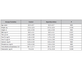
Гіпертиреоз є фактором ризику зниження мінеральної щільності кісткової тканини (МЩКТ) та виникнення остеопоротичних переломів. Оцінка вертебральних переломів (ОВП) допомагає діагностувати вертебральний перелом, а показник якості трабекулярної кісткової тканини (ТКТ) — оцінити кісткову мікроархітектуру. Ми поставили собі за мету оцінити вплив гіпертиреозу на МЩКТ, склад м’яких тканин тіла, частоту «мовчазних» (невиявлених) вертебральних переломів і ТКТ. 80 пременопаузальних португальських жінок були розподілені в групи пацієнтів з підтвердженим гіпертирео­зом (n = 40) і контрольну групу (n = 40). МЩКТ (г/см2) поперекового відділу хребта, стегнової кістки, 33 % променевої кістки, усього скелета досліджували за допомогою двохенергетичної рентгенівської абсорбціометрії (ДРА) з урахуванням маси тіла (кг) пацієнтів. ОВП застосовувалась для діагностики переломів, що згодом класифікували за напівкількісним методом Женанта та підтверджували за допомогою рентгенографії. Показник ТКТ отримували за допомогою ДРА поперекового відділу хребта. Жоден пацієнт раніше не отримував лікування щодо гіпертиреозу, остеопорозу або зниженої кісткової маси (остеопенії). Отримані результати підтверджувалися статистичними підрахунками. У групі пацієнтів з гіпертиреозом середня МЩКТ (МЩКТ стегнової кістки, шийки стегнової кістки, усього скелета), загальна знежирена маса та ТКТ були вірогідно нижчими, ніж у контрольній групі; згідно з класифікацією Міжнародної асоціації клінічної денситометрії (ISCD), відзначалася тенденція до більшої частоти зниження МЩКТ; кількість вертебральних переломів також вірогідно зростала. Ці результати свідчать про те, що в групі пременопаузальних жінок з гіпертиреозом показники МЩКТ, знежиреної маси та ТКТ є вірогідно нижчими. Частота «мовчазних» вертебральних переломів є також вірогідно вищою.

Гипертиреоз является фактором риска снижения минеральной плотности костной ткани (МПКТ) и возникновения остеопоротических переломов. Оценка вертебральных переломов (ОВП) помогает диагностировать вертебральный перелом, а показатель качества трабекулярной костной ткани (ТКТ) — оценить костную микроархитектуру. Мы поставили себе цель оценить влияние гипертиреоза на МПКТ, состав мягких тканей тела, частоту «немых» (невыявленных) вертебральных переломов и ТКТ. 80 пременопаузальных португальских женщин были распределены в группу пациентов с подтвержденным гипертиреозом (n = 40) и контрольную группу (n = 40). МПКТ (г/см2) поясничного отдела позвоночника, бедренной кости, 33 % лучевой кости, всего скелета исследовали с помощью двухэнергетической рентгеновской абсорбциометрии (ДРА) с учетом массы тела (кг) пациентов. ОВП применялась для диагностики переломов, которые впоследствии классифицировали с применением полуколичественного метода Женанта и подтверждали с помощью рентгенографии. Показатель ТКТ получали с помощью ДРА поясничного отдела позвоночника. Ни один пациент ранее не получал лечения по поводу гипертиреоза, остеопороза или пониженной костной массы (остеопении). Полученные результаты подтверждались статистическими подсчетами. В группе пациентов с гипертиреозом средняя МПКТ (МПКТ бедренной кости, шейки бедренной кости, всего скелета), общая обезжиренная масса и ТКТ были достоверно ниже, чем в контрольной группе; согласно классификации Международной ассоциации клинической денситометрии (ISCD), отмечалась тенденция к большей частоте снижения МПКТ; количество вертебральных переломов также достоверно возрастало. Эти результаты свидетельствуют о том, что в группе пременопаузальных женщин с гипертиреозом показатели МПКТ, обезжиренной массы и ТКТ являются достоверно более низкими. Частота «немых» вертебральных переломов также достоверно выше.

Hyperthyroidism is a risk factor for reduced bone mineral density (BMD) and osteoporotic fractures. Vertebral fracture assessment (VFA) detects vertebral fractures and trabecular bone score (TBS) is an index of bone microarchitecture. We aimed to evaluate the effects of hyperthyroidism on BMD, soft body tissue composition, prevalence of silent vertebral fractures and TBS. Eighty Portuguese premenopausal women were divided and matched in overt hyperthyroidism (n = 40) and control (n = 40) groups. BMD (g/cm2) at lumbar spine, hip, radius 33 % and whole body and the total body masses (kg) were studied by DXA. VFA was used to detect fractures and those were classified by Genant’s semiquantitative method and confirmed by X-ray. TBS was obtained from lumbar spine DXA images. No patient had previously been treated for hyperthyroidism, osteoporosis or low bone mass. Adequate statistical tests were used. In the hyperthyroidism group, the mean BMD (total hip, femoral neck and whole body), the total lean mass and TBS were significantly lower; according to ISCD classification, there was a trend for a higher prevalence of low BMD; vertebral fractures were significantly higher. These results suggest that in a group of hyperthyroid premenopausal women there are significantly lower BMD, lean mass and TBS. The prevalence of silent vertebral fractures is also significantly higher.


Ключевые слова

показник якості трабекулярної кісткової тканини; остеопороз; гіпертиреоз; переломи; пременопаузальні жінки

показатель качества трабекулярной костной ткани; остеопороз; гипертиреоз; переломы; пременопаузальные женщины

TBS; osteoporosis; hyperthyroidism; vertebral fractures; premenopausal women

Introduction

It is known that the excess of circulating thyroid hormones can lead to an increase of bone resorption, either by acting directly on osteoclasts or indirectly on osteoblasts [1]. Also thyroid stimulating hormone (TSH) seems to be a negative regulator of bone remodeling, inhibiting the formation, the survival of osteoclasts and the differentiation of osteoblasts [2, 3]; however, this effect has not been totally clarified because experiments in mice with a loss-of-function TSH receptor, the bone loss seems to be independent of TSH levels [4]. 
In a clinical point of view, overt hyperthyroidism has been recognized to be an important cause of secondary osteoporosis and a risk factor for hip fracture in women; osteoporotic fractures are associated with a risk of precocious mortality, namely in the elderly [5].
Moreover, several studies proposed that low TSH levels, per se, can predispose to osteoporosis and fragility fractures [6–8], but this subject is far from clarified; most of the studies are based on the effect of supraphysiological doses of thyroid hormone to suppress TSH secretion in the treatment of differentiated thyroid carcinoma or nontoxic goiter in postmenopausal women and still provide conflicting results [9]. However, the clinical studies addressing the effects of hyperthyroidism on the skeleton in young and premenopausal women are still scarce.
A previous study by our group in hyperthyroid men aged less than 50 years, showed bone mineral density (BMD) decreases in all skeletal regions (non-significant just at the distal radius) and an increase in the prevalence of vertebral fractures detected by Vertebral fracture assessment (VFA) [10].
Vertebral fractures are among the most common in osteoporosis. Their diagnosis is important because it predicts the occurrence of future fragility fractures all over the skeleton [11]. Moreover, around 69 % of patients with vertebral fractures are unaware of them, not only due to the absence of symptoms but also because patients are not routinely or accurately imaged. They occur more frequently in patients known to have low bone mass more than osteoporosis diagnosis by dual x-ray absorptiometry (DXA) [12]. VFA by DXA is a spine imaging with DXA scanners and may represent a better alternative to conventional radiography in the diagnosis of vertebral fractures due to lower radiation dose and also to greater convenience for the patient as it can be done at the same time of DXA [13].
The trabecular bone score (TBS) is a relatively recent and indirect index of bone microarchitecture which can improve the prediction of fracture risk not only in osteoporosis but also in some metabolic bone diseases. The experimental variogram method is used to estimate the bone microarchitecture. An increased TBS value correlates with better bone microarchitecture, while a reduced one correlates with a fragile bone microarchitecture. It was shown that TBS is associated with the structure of bone tissue and it may detect differences between DXA scans that show identical BMD amounts. So, in addition to clinical risk factors and BMD, TBS can be even more sensitive than BMD in identifying secondary osteoporosis like in cases of hyperparathyroidism, adrenal adenomas and iatrogenic reasons. However, in hyperthyroidism, clinical studies with TBS data are very scarce [14, 15].
The aims of our study were to evaluate the effects of overt hyperthyroidism in premenopausal women on: BMD, soft body tissue composition, prevalence of silent vertebral fractures detected by VFA and TBS. Its scientific relevance is due to the fact that clinical studies addressing these effects, namely data about fracture susceptibility and TBS are relatively scarce, especially in premenopausal women.

Materials and methods

Eighty premenopausal women were divided and matched in the hyperthyroidism group (n = 40) and in the control group (n = 40). From 50 initial patients with hyperthyroidism which were referred to the Endocrinology Department for diagnosis and treatment, only 40 were recruited for the study. For each patient, an age (limits 6–11 months) and stature (limits 1–3 cm) matched control person was drawn from a random sample of the endocrinology clinic patients without diseases affecting bone metabolism. Exclusion criteria for both patients and controls were: hypo/hyperparathyroidism, hypogonadisms, diabetes mellitus, hypo/hypercortisolism, vitamin D deficiency, inflammatory bowel disease, malabsorption diseases, liver/renal diseases, neoplasia, and medications affecting the skeleton including L-thyroxine.
Regarding the etiology of the hyperthyroidism, 33 cases were Graves’s disease and 7 cases were toxic nodular goiter.
No patient had previously been treated empirically for osteoporosis or reduced bone mass or hyperthyroidism. We cannot be sure of the duration of hyperthyroidism before the beginning of anti-thyroid medication.
Also, past history of fragility fractures and symptoms of vertebral fracture were excluded in both patients and controls. All patients and controls had a full clinical examination and BMI (kg/m2) was calculated.
In both groups, BMD (g/cm2) at the lumbar spine, at the hip (femoral neck and total), at the distal radius (1/3 or 33 %) and at the whole body and total body tissue composition including soft body lean and fat masses (kg) were studied by DXA using the QDR Discovery W radiological densitometer (Hologic Inc.) of the Lisbon Clinic of Endocrinology Diabetes and Metabolism, Lda. (CEDML).
According to ISCD recommendations, in both groups BMD was qualified by the lowest Z-score obtained at the lumbar spine, at the hip and at the distal radius (33 %) in low and normal BMD [16].
TBS was obtained from the pixel grey-level texture metric analysis of the two-dimensional lumbar spine DXA images (iNsight software TM version 3.0.2.0, Medimaps, Mérignac, France) [14].
We analyzed the percentage of patients and controls in the 3 groups of TBS qualification: higher or equal to 1.310 — high TBS, low fracture risk; between 1.230 and 1.310 — medium TBS, medium fracture risk; less or equal to 1.230 — low TBS, high fracture risk [17, 18].
The lateral images of thoracolumbar spine in DXA scan (VFA) were used to detect fractures and those were classified according to type (wedge, biconcave, crush) and severity (% of deformity) by Genant’s Semiquantitative Method, by one endocrinologist. This method combines the qualitative visualization of the spine with the morphometric measurements of the vertebral body height in 6 points [19].
In order to more precisely confirm the fractures, all patients had thoracolumbar spine X-ray (on frontal and lateral projections) on the same day or within a few days, which was reviewed by one radiologist. In a few instances where there was disagreement, a second radiologist was consulted. Conventional radiographs were electronic images produced by digital X-ray equipment and were viewed using a high-resolution viewing workstation designed for medical image reading. Their final reports were established as the gold standard for proven vertebral fractures, and only the positive cases in both VFA and X-ray were considered.
Fasting blood samples were collected for measurement of serum chemistries and blood counts, hormones and osteocalcin. Serum free T3, free T4 and TSH were assayed by an electrochemiluminescence immunoassay (Roche) and total calcium and phosphorus were assayed by enzymatic colorimetry (Roche).
The study was approved by Santa Maria University Hospital (HSM-CHLN, EPE) and Lisbon Medical School Faculty of Medicine of the Lisbon University (FMUL) with the IRB approval nº HSM-CHLN, EPE/FMUL nº 890/10.
All patients and controls gave their informed consent, according to the approved protocol by the ethic committee of the institution and based on Helsinki declaration.
Statistical Methods: the data were statistically analyzed using the Statgraphics Centurion XVI version 16.1.07.01. All the results are expressed as mean ± SD. After testing for normal distribution, the Chi-square test was used to compare the differences in parametric data between groups. The Fisher’s exact test was used to compare the number of fractures in both groups. We also performed correlation analysis. P value < 0.05 was considered statistically significant.

Results

The mean age, height and mean total body fat mass were similar between the groups. In the hyperthyroidism group, the serum concentrations of alkaline phosphatase and osteocalcin were significantly higher compared to controls (Table 1).
In the hyperthyroidism group, the mean lean mass was significantly lower, while the BMD was also lower in all the skeletal regions, reaching significance at total hip, femoral neck and whole body (Table 2). In the same group, the prevalence of low BMD was higher, but not significantly.
In the control group we found 2 patients with fractures (5 %) both grades 1 and 2. In the hyperthyroidism group (n = 40) we found 8 patients with fractures (8/40) (Table 3 and Figure 1): 2 fractures of grade 1 in 5 patients and fractures of both grades 1 and 2 in 3 patients. All of them were confirmed by conventional X-ray. BMD qualification of the women with fractures, was: control group — low BMD; hyperthyroidism group — normal BMD in 2, low BMD in 6. All the fractures except 1, were localized in the thoracic spine. 
The mean TBS in both groups were in the considered normal range (Table 2), however the mean TBS in hyperthyroid patients was significantly lower than in controls (1.406 ± 0.08 vs 1.463 ± 0.08 respectively; p = 0.005). Moreover, when we analyzed the percentage of patients and controls in the 3 groups of TBS qualification (Table 4), no one was found in the high fracture risk (TBS < 1.230), while a significantly higher percentage of hyperthyroid patients was found in the medium fracture risk (TBS ≥ 1.230 and < 1.310). TBS values were similar between the women with and without fractures in both groups.

Discussion

In adults, the excessive levels of thyroid hormones in blood may originate bone loss. 
Clinical studies addressing the bone consequences of hyperthyroidism (both overt and subclinical), namely BMD and fragility fractures, are relatively scarce, mainly in young populations.
The importance of our study is due to the fact that it was done in a population of premenopausal women, with overt hyperthyroidism naïve of treatment and we found a decreased BMD in all skeletal regions, with significance at total hip, femoral neck and whole body.
Several clinical studies addressing the negative effects of thyroid hormones on bone have described it in all the skeleton (both axial and appendicular), however more pronounced in areas consisting mainly of cortical bone like the femoral neck and the distal radius [20–22].
A cross-sectional study of premenopausal women, found significantly low BMD at several skeletal regions, in hyperthyroid women compared to controls, which did not appear to be associated to the duration of the disease (except for distal radius) [23].
In our study, the low BMD at both lumbar spine and distal radius but not reaching statistical significance could be explained by the small number of patients. 
Regarding the etiology of hyperthyroidism, our patients had mainly Graves’s disease (33 cases) and nodular goiter (7 cases). Besides it is not totally clarified, these etiologies do not seem to influence the negative consequences on BMD; however, it increases the prevalence of osteoporosis [24]. In our study, there were no differences regarding BMD, but there was a tendency for a low (non-significant) total lean mass in patients with Graves’s disease compared to those with toxic goiter, probably because of the more prolonged and severe hyperthyroidism.
The significantly low total body lean mass (not the total fat mass), weight and BMI observed in the hyperthyroidism group as compared to the control group can be explained by the weight loss and gastrointestinal changes (increased gut motility, diarrhea and consequent malabsorption of proteins, minerals and vitamins) associated with hyperthyroidism and all those factors together may also provoke bone mass loss.
In the hyperthyroidism group we found correlations of the weight and of the BMI with both total femur BMD and femoral neck BMD, but we also found those correlations in the control group. We did not find correlations of the TSH nor the free T3/T4 with the BMD, which can be explained by the variability in the duration of the active non-treated hyperthyroidism phase.
So, our study was not designed to answer if the mechanism of bone loss was mainly the weight/BMI/total lean and fat masses reductions or the thyroid hormones/decrease TSH bone actions, but we believe that both were implicated. 
The Rotterdam study, done in a large sample of elderly Caucasian men and women, suggested that besides the effect of weight and BMI on bone density, there is also a direct effect of thyroid function (namely TSH and free T4) on bone tissue [25].
Vertebral fractures are the most common fractures in osteoporosis but most of them are not recognized and not diagnosed. It is known that about 10 to 28 % of women over 65 years-old has one or more prevalent vertebral fractures.
The data about osteoporotic fractures risk, namely vertebral fractures, in a specifically young female population with endogenous hyperthyroidism are relatively scarce.
A meta-analysis from P. Vestergaard et al. showed that the risk of hip fractures increased significantly with age at diagnosis of hyperthyroidism [26]. A more recent meta-analysis from M. Blum et al. in endogenous subclinical hyperthyroidism (TSH < 0.1 mU/L) showed vertebral fractures in 1.3 % in 6 studies with a hazard ratio for vertebral fractures of 1.74 (95% CI, 1.01–2.99) [27].
Several other clinical studies were done in older populations and with iatrogenic hyperthyroidism, and showed that fracture risk (hip and vertebral) was higher in older men and mainly in women with a suppressed TSH [28–30]. However, it is not clear that iatrogenic hyperthyroidism does affect bone in a totally similar way as hyperthyroidism due to toxic goiter or autoimmune diseases.
So, in our study, the decreases in both bone and total lean masses in the hyperthyroid women were probably important factors leading to the increase in fracture risk observed in this population, but other factors were also probably implicated.
Clinical studies of TBS in premenopausal women are scarce and in non iatrogenic hyperthyroid premenopausal women are even scarcer. 
In our study, the mean TBS in the control and in the hyperthyroidism groups were in the normal range, but the mean TBS in hyperthyroid patients was significantly lower than in the control group.
Moreover, when we analyzed the percentage of patients and controls in the 3 groups of TBS qualification, we did not find any control person or hyperthyroid patient in the high fracture risk; nevertheless, a higher percentage of hyperthyroid patients were detected in the medium fracture risk. 
S.Y. Ock et al found in Graves’s disease male and female patients a significant increase of TBS values from 1.377 to 1.390 after anti-thyroid therapy, however it was a non-controlled study and those scores are considered within normal range [31].
In a cross-sectional controlled study of premenopausal women with Graves’ disease, Kuzma et al. found that TBS levels were lower in active and cured patients compared to controls, showing a strong correlation of the disease with decreased TBS [32].
Comparing the patients with and without fractures in both groups, there were no differences in the TBS. A possible explanation is that the cortical bone is usually more and precociously affected than the trabecular bone (which is evaluated by TBS), in this endocrine disease. Another explanation could be that the time with non-treated hyperthyroidism was not long enough to develop bone microarchitecture changes measured by TBS.
The main limitation of our study was the relatively small number of patients.
The strength of our study was the fact of being done in a population of premenopausal women not previously treated for hyperthyroidism, osteoporosis or low bone mass and already had lower lean mass and BMD, as well as more vertebral fractures when compared to controls. Moreover, to our knowledge, this is the first study of TBS data in hyperthyroid premenopausal women. 
However, future studies with bigger female populations and also evaluating the effects of anti-thyroid treatment will be important to better understand the bone disease of hyperthyroidism.

Conclusions

In this controlled study of premenopausal women with overt hyperthyroidism, we found significantly lower BMD (mainly cortical bone BMD), total lean body mass and TBS. We also found a higher prevalence of silent vertebral fractures detected by VFA technology and confirmed by X-ray.
Conflicts of interests. Authors declare the absence of any conflicts of interests and their own financial interest that might be construed to influence the results or interpretation of their manuscript.
Funding. This research did not receive any specific grant from any funding agency in the public, commercial or not-for-profit sector.

Список литературы

1. Britto J.M., Fenton A.J., Holloway W.R., Nicholson G.C. Osteoblasts mediate thyroid hormone stimulation of osteoclastic bone resorption. Endocrinology. 1994. 134. 169-176.

2. Abe E., Marians R.C., Yu W. TSH is a negative regulator of skeletal remodeling. Cell. 2003. 115(2). 151-162.

3. Sun L., Zhu L.L., Lu P. Genetic confirmation for a central role for TNFα in the direct action of thyroid stimulating hormone on the skeleton. Proc. Natl. Acad. Sci USA. 2013. 110. 9891-9896.

4. Bassett J.H., O’Shea P.J., Sriskantharajah S. Thyroid hormone excess rather than thyrotropin deficiency induces osteoporosis in hyperthyroidism. Mol. Endocrinol. 2007. 21. 1095-1107.

5. Cummings S.R., Nevitt M.C., Browner W.S. Risk factors for hip fracture in white women. Study of Osteoporotic Fractures Research Group. N. Engl. J. Med. 1995. 332. 767-773.

6. Mudde A.H., Reijnders F.J.L., Nieuwenhuijzen A.C. Peripheral bone density in women with untreated multinodular goitre. Clin. Endocrinol. 1992. 37. 35-39.

7. Földes J., Tarján G., Szathmari M. Bone mineral density in patients with endogenous subclinical hyperthyroidism: Is this thyroid status a risk factor for osteoporosis? Clin. Endocrinol. 1993. 39. 521-527.

8. Mazziotti G., Porcelli T., Patelli I. Serum TSH values and risk of vertebral fractures in euthyroid post-menopausal women with low bone mineral density. Bone. 2010. 46(3). 747-751.

9. Baran D. Thyroid Hormone and the Skeleton. In: Marcus R., Feldman D. (eds.). Osteoporosis. 4th ed. Elsevier, Oxford. 2013. 1239-1247.

10. Barbosa A.P., Mascarenhas M.R., Silva C.F. Prevalence of silent vertebral fractures detected by vertebral fracture assessment in young Portuguese men with hyperthyroidism. Eur. J. Endocrinol. 2015. 172. 189-194.

11. Black D.M., Arden N.K., Palermo L. Prevalent Vertebral deformities predict hip fractures and new vertebral deformities but not wrist fractures. J. Bone Miner. Res. 1999. 14(5). 821-828.

12. Jager P., Jonkman S., Koolhaas W. Combined vertebral fracture assessment and bone mineral density measurement: a new standard in the diagnosis of osteoporosis in academic populations. Osteoporos Int. 2011. 22(4). 1059-1068.

13. Rea J.A., Chen M.B., Li J. Morphometric X-ray absorptiometry and morphometric radiography of the spine: a comparison of prevalent vertebral deformity identification. J. Bone Miner. Res. 2000. 15. 564-574.

14. Bousson V., Bergot C., Sutter B. Trabecular bone score (TBS): available knowledge, clinical relevance, and future prospects. Osteoporos Int. 2012. 23(5). 1489-1501.

15. Silva B.C., Leslie W.D., Resch H. Trabecular Bone Score: A Noninvasive Analytical Method Based Upon the DXA Image. J. Bone Miner. Res. 2014. 29(3). 518-530.

16. Schousboe J.T., Shepherd J.A., Bilezikian J.P. Executive Summary of the 2013 ISCD Position Development Conference on Bone Densitometry. J. Clin. Densitom. 2013. 16(4). 455-467.

17. McClosky E.V., Odén A., Harvey N.C. A Meta-Analysis of Trabecular Bone Score in Fracture Risk Prediction and its Relationship to FRAX. J. Bone Min. Res. 2016. 31(5). 940-948.

18. Medimaps Group. 2016 TBS Reference Curves.

19. Genant H.K., Wu C.Y., van Kuijk C. Vertebral fracture assessment using a semiquantitative technique. J. Bone Miner. Res. 1993. 8. 1137-1148.

20. Van den ven A.C., Erdtsieck R.J. Changes of bone mineral density, quantitative ultrasound parameters and markers of bone turnover during treatment of hyperthyroidism. The Netherlands J. Medicine. 2008. 66(10). 428-432.

21. Greenspan S.L., Greenspan F.S. The effect of thyroid hormone on skeletal integrity. Annals of Internal Medicine. 1999. 130(9). 750-758.

22. Langdahl B.L., Loft A.G., Eriksen E.F. Bone mass, bone turnover, calcium homeostasis, and body composition in surgically and radioiodine-treated former hyperthyroid patients. Thyroid. 1996. 6. 169-175.

23. Boonya-Ussadorn T., Punkaew B., Sriassawaamorn N. A comparative study of bone mineral density between premenopausal women with hyperthyroidism and healthy premenopausal women. J. Med. Associat. Thailand. 2010. 93(suppl. 6). S1-S5.

24. Svare A., Nilsen T.I., Bjøro T. Hyperthyroid levels of TSH correlate with low bone mineral density: the HUNT 2 study. Eur. J. Endocrinol. 2009. 161. 779-786.

25. Van der Deure W.M., Uitterlinden A.G., Hofman A. Effects of serum TSH and FT4 levels and the TSHR-Asp727Glu polymorphism on bone: the Rotterdam Study. Clin. Endocrinol. (Oxf.). 2008. 68. 175-181.

26. Vestergaard P., Mosekilde L. Hyperthyroidism, bone mineral and fracture risk — a meta-analysis. Thyroid. 2003. 13(6). 585-93.

27. Blum M., Bauer D., Collet T. Subclinical thyroid dysfunction and fracture risk a meta-analysis. JAMA. 2015. 313(20). 2055-65.

28. Lee J.S., Buzková P., Fink H.A. Subclinical thyroid dysfunction and incident hip fracture in older adults. Arch. Intern. Med. 2010. 170. 1876-1883.

29. Heijckmann A.C., Huijberts M.S.P., Geusens P. Hip bone mineral density, bone turnover and risk of fracture in patients on long-term suppressive L-thyroxine therapy for differentiated thyroid carcinoma. Eur. J. Endocrinol. 2005. 153. 23-29.

30. Turner M.R., Camacho X., Fischer H.D. Levothyroxine dose and risk of fractures in older adults: nested case-control study. British. Med. J. 2011. 342. 1-9.

31. Ock S.Y., Chung Y., Choi Y.J. Changes in bone mineral density and trabecular bone score in Graves’ disease patients after anti-thyroid therapy. Osteoporosis and Sarcopenia. 2016. 2. 175-179.

32. Kuzma M., Vanuga P., Binkley N. High serum fractalkine is associated with lower trabecular bone score in premenopausal women with Graves’s disease. Horm. Metab. Res. 2018. 50(8). 609-614.


Вернуться к номеру