
Журнал «Боль. Суставы. Позвоночник» Том 15, №2, 2025
Вернуться к номеру
Варіації положення надколінка залежно від морфотипу колінного суглоба у хворих з остеоартритом
Авторы: Килимнюк Л.О. (1), Маціпура М.М. (2), Яремин С.Ю. (2)
(1) - Медичний центр «Angels Clinic», м. Вінниця, Україна
(2) - Вінницький національний медичний університет ім. М.І. Пирогова, м. Вінниця, Україна
Рубрики: Ревматология, Травматология и ортопедия
Разделы: Клинические исследования
Версия для печати
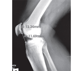
Актуальність. Вивчення особливостей положення надколінка з урахуванням морфотипу колінного суглоба є актуальним завданням сучасної ортопедії. Мета: охарактеризувати особливості положення надколінка у хворих з остеоартритом колінного суглоба з урахуванням морфологічних варіантів структури суглоба. Матеріали та методи. Проаналізовано результати рентгенографічного обстеження 100 випадків остеоартриту колінного суглоба (середній вік обстежених — 63,56 ± 8,10 року). Морфотип І встановлено у 21 (21,0 %) пацієнта, морфотип ІІ — у 38 (38,0 %), морфотип ІІІ — у 29 (29,0 %), морфотип IV — у 12 осіб (12,0 %). Особливості положення надколінка визначали, розраховуючи індекси Insall-Salvati, Caton-Deschamps, Grelsamer-Meadows, Blackburne-Peel. Рівень статистичної значущості визначали при р ≤ 0,05. Результати. Середній показник індексу Insall-Salvati в осіб з морфотипом І становив 1,11 ± 0,05, морфотипом ІІ — 1,29 ± 0,38, морфотипом ІІІ — 1,32 ± 0,25, морфотипом ІV — 1,40 ± 0,14 (р = 0,009). Значення індексу Caton-Deschamps в обстежених з морфотипом І становило 0,77 ± 0,14, морфотипом ІІ — 0,74 ± 0,23, морфотипом ІІІ — 0,78 ± 0,24, морфотипом ІV — 1,05 ± 0,16 (р = 0,02). Середній показник індексу Grelsamer-Meadows в обстежених з морфотипом І становив 1,84 ± 0,12, морфотипом ІІ — 1,86 ± 0,46, морфотипом ІІІ — 1,86 ± 0,44, морфотипом ІV — 2,41 ± 0,33 (р = 0,009). Показник відношення Blackburne-Peel у пацієнтів з морфотипом І становив 0,64 ± 0,09, морфотипом ІІ — 0,59 ± 0,23, морфотипом ІІІ — 0,74 ± 0,24, морфотипом ІV — 0,98 ± 0,14 (р = 0,001). Наявність значень Blackburne-Peel < 0,8 була асоційована з вищими шансами формування морфотипу ІІ (відношення шансів (OR) = 9,8; 95% CI: 1,9–49,2; p = 0,0007). Вищі шанси розвитку морфотипу ІІІ встановлено у пацієнтів зі значеннями Insall-Salvati > 1,2 (OR = 4,7; 95% CI: 1,4–16,0; p = 0,007), Blackburne-Peel 0,8–1,0 (OR = 4,4; 95% CI: 1,2–17,0; p = 0,03). Наявність значень Caton-Deschamps > 1,2 та Blackburne-Peel > 1,0 була пов’язана з вищими шансами формування морфотипу ІV (OR = 21,6; 95% CI: 1,6–297,4; p = 0,02, та OR = 23,1; 95% CI: 3,3–160,1; p = 0,001, відповідно). Висновки. Особливості положення надколінка у пацієнтів з морфотипами І та ІІ колінного суглоба зумовлені переважно патологічними змінами суглобової поверхні проксимального відділу великогомілкової кістки, у хворих з морфотипами ІІІ та IV — ураженням пателофеморального зчленування.
Background. Patellar position relative to knee joint morphotype is clinically relevant. The purpose was to characterize patellar position features in patients with knee osteoarthritis, considering morphological joint variants. Materials and methods. Radiographs of 100 patients (mean age 63.56 ± 8.10 years) with medial knee osteoarthritis were analyzed. Morphotype I was identified in 21.0 %, morphotype II in 38.0 %, morphotype III in 29.0 %, morphotype IV in 12.0 % of cases. Patellar position was assessed using Insall-Salvati, Caton-Deschamps, Grelsamer-Meadows, Blackburne-Peel indices. Statistical significance was set at p ≤ 0.05. Results. Mean Insall-Salvati index was 1.11 ± 0.05 in morphotype I, 1.29 ± 0.38 in morphotype II, 1.32 ± 0.25 in morphotype III, 1.40 ± 0.14 in morphotype IV (p = 0.009). Caton-Deschamps index was 0.77 ± 0.14 in morphotype I, 0.74 ± 0.23 in morphotype II, 0.78 ± 0.24 in morphotype III, and 1.05 ± 0.16 in morphotype IV (p = 0.02). Mean Grelsamer-Meadows index was 1.84 ± 0.12 in morphotype I, 1.86 ± 0.46 in morphotype II, 1.86 ± 0.44 in morphotype III, and 2.41 ± 0.33 in morphotype IV (p = 0.009). Blackburne-Peel ratio was 0.64 ± 0.09 in morphotype I, 0.59 ± 0.23 in morphotype II, 0.74 ± 0.24 in morphotype III, and 0.98 ± 0.14 in morphotype IV (p = 0.001). Blackburne-Peel index < 0.8 was associated with a higher likelihood of morphotype II (OR = 9.8; 95% CI: 1.9–49.2; p = 0.0007). Increased odds of morphotype III were associated with Insall-Salvati index > 1.2 (OR = 4.7; 95% CI: 1.4–16.0; p = 0.007), Blackburne-Peel 0.8–1.0 (Odd Ratio (OR) = 4.4; 95% CI: 1.2–17.0; p = 0.03). Caton-Deschamps > 1.2 and Blackburne-Peel > 1.0 were associated with greater odds of morphotype IV (OR = 21.6; 95% CI: 1.6–297.4; p = 0.02, and OR = 23.1; 95% CI: 3.3–160.1; p = 0.001, — respectively). Conclusion. Therefore, in morphotypes I and II, patellar position alterations were primarily associated with pathological changes in the proximal tibia, whereas in morphotypes III and IV, they were predominantly linked to patellofemoral joint involvement.
колінний суглоб; дегенеративно-дистрофічні захворювання; остеоартрит; гонартроз; надколінок; морфологія
knee joint; degenerative-dystrophic diseases; osteoarthritis; gonarthrosis; patella; morphology
Для ознакомления с полным содержанием статьи необходимо оформить подписку на журнал.
- Konrads C, Schreiner AJ, Cober S, Schüll D., Ahmad SS, Alshrouf MA. Evaluation of patella height in native knees and arthroplasty: an instructional review. SICOT-J. 2022;8:36. doi: 10.1051/sicotj/2022037.
- Analan PD, Ozdemir H. The Effect of Patellar Height by Using Insall Salvati Index on Pain, Function, Muscle Strength and Postural Stability in Patients with Primary Knee Osteoarthritis. Current Medical Imaging. 2021;17(4):532-538. doi: 10.2174/1573405616999200817172649.
- D’Ambrosi R, Rubino F, Ursino C, et al. Change in patellar height in medial and lateral unicompartmental knee arthroplasty: a clinical trial. Archives of Orthopaedic аnd Trauma Surgery. 2024;144(3):1345-1352. doi: 10.1007/s00402-023-05139-8.
- Lum ZC, Saiz AM, Pereira GC, Meehan JP. Patella Baja in Total Knee Arthroplasty. The Journal of the American Academy of Orthopaedic Surgeons. 2020;28(8):316-323. doi: 10.5435/JAAOS-D-19-00422.
- Lee H, Fletcher C, Hartwell M, Strickland SM. Effect of Patellofemoral Arthroplasty on Patellar Height in Patients with Patellofemoral Osteoarthritis. The Journal оf Knee Surgery. 2023;36(12):1283-1288. doi: 10.1055/s-0042-1755354.
- Biedert RM. Patella Alta: When to Correct and Impact on Other Anatomic Risk Factors for Patellofemoral Instability. Clinics in Sports Medicine. 2022;41(1):65-76. doi: 10.1016/j.csm.2021.07.002.
- D’Ambrosi R, Buda M, Nuara A, et al. Patellar height after unicompartmental knee arthroplasty: comparison between fixed and mobile bearing. Archives of Orthopaedic аnd Trauma Surgery. 2022;142(11):3449-3460. doi: 10.1007/s00402-021-04183-6.
- Schreiner AJ, Spiegel L, Yan SG, et al. Evaluation of modified and newly applied patella height indices in primary total knee arthroplasty. Skeletal Radiology. 2023;52(1):73-82. doi: 10.1007/s00256-022-04142-1.
- Cassar-Pullicino VN, Davies AM, еditors. Measurements in Musculoskeletal Radiology. Berlin, Germany: Springer; 2020. 860 p.
- Insall J, Salvati E. Patella position in the normal knee joint. Radiology. 1971;101(1):101-104. https://doi.org/10.1148/101.1.101.
- Grelsamer RP, Meadows S. The modified Insall-Salvati ratio for assessment of patellar height. Clinical Orthopaedics аnd Related Research. 1992;(282):170-176.
- Blackburne JS, Peel TE. A new method of measuring patellar height. The Journal of Bone аnd Joint Surgery. British volume. 1977;59(2):241-242. doi: 10.1302/0301-620X.59B2.873986.
- Caton J, Deschamps G, Chambat P, Lerat JL, Dejour H. Les rotules basses. A propos de 128 observations [Patella infera. Apropos of 128 cases]. Revue de Chirurgie Orthopedique еt Reparatrice de L’appareil Moteur. 1982;68(5):317-325.
- Konrads C, Rejaibia J, Grosse LC, et al. Patella-height analysis and correlation with clinical outcome after primary total knee arthroplasty. Journal of Оrthopaedics. 2021;23:169-174. doi: 10.1016/j.jor.2021.01.001.
- Suthar A, Yukata K, Azuma Y, et al. Significant reduction of patellar height affected lower clinical outcomes and knee flexion over five-year follow-up after total knee arthroplasty. Bone & Joint Open. 2021;2(12):1075-1081. doi: 10.1302/2633-1462.212.BJO-2021-0164.R1.
- Verhulst FV, van Sambeeck JDP, Olthuis GS, van der Ree J, Koëter S. Patellar height measurements: Insall-Salvati ratio is most reliable method. Knee Surgery, Sports Traumatology, Arthroscopy: official journal of the ESSKA. 2020;28(3):869-875. doi: 10.1007/s00167-019-05531-1.
- Kylymniuk LO, Matsipura MM, Iaremyn SIu. Clustering of Knee Joint Types Affected by the Degenerative-Dystrophic Process Clustering of Knee Joint Types Affected by the Degenerative-Dystrophic Process. Укр. мед. часопис. 2025;2(168):1-6. doi: 10.32471/umj.1680-3051.264438. [article in English].
- Sahanand KS, Pandian P, Chellamuthu G, Rajan DV. Effect of ascending and descending medial open wedge high tibial osteotomy on patella height and functional outcomes — a retrospective study. European Journal оf Orthopaedic Surgery & Traumatology: Orthopedie Traumatologie. 2024;34(1):499-505. doi: 10.1007/s00590-023-03693-w.
- Kudo Y, Maeyama A, Ishimatsu T, et al. Changes in patellar baja progress until 3 months after medial open-wedge high tibial osteotomy. Journal of Orthopaedic Surgery (Hong Kong). 2022;30(3):10225536221128615. doi: 10.1177/10225536221128615.
- Luceri F, Basilico M, Batailler C, et al. Effects of sa–gittal tibial osteotomy on frontal alignment of the knee and patellar height. International Оrthopaedics. 2020;44(11):2291-2298. doi: 10.1007/s00264-020-04580-3.
- Sim JA, Na YG, Lee BK, Lee BH. Alignment changes after open-wedge high tibial osteotomy result in offloa–ding in the patellofemoral joint: a SPECT/CT analysis. Knee Surgery, Sports Traumatology, Arthroscopy: official journal of the –ESSKA. 2022;30(2):437-446. doi: 10.1007/s00167-020-06115-0.